Small Molecular Analytical Lab
Department of Biotechnology has the following facilities for analyzing characteristic small molecules from complex media or mixtures. The small molecular analytical lab is designed for such discoveries and to make use of the findings in developing new, more efficient, easy to perform diagnostic procedure on a variety of microbial pathogen.
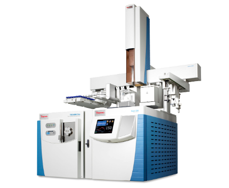
- The GC/MS can be used for the bioanalysis of body fluids to detect narcotics, barbiturates, alcohols, and drugs such as anticonvulsants, anaesthetics, antihistamines, sedative hypnotics, and anti-epileptic drugs. It is also useful in detecting pollutants and metabolites in serum and in fatty acid profiling in microbes
- Gas chromatography (GC) is normally the preferred technique for the determination of impurities in solvents
- “Due to the reduced downtime of the TSQ 9610, more samples can be evaluated, which generates a higher sample throughput per employee resulting in a higher analytical efficiency
- “Due to the extended dynamic range of the TSQ 9610 GC-MS/MS, high-level PCBs and low-level dioxins can be quantified in a single method also increased throughput due to the robustness of the instrument and its ability to handle dirty matrices such as soil, industrial smokestack samples, and wastewater This ensures we do not have to re-clean and re-run samples, which saves us time. This enabled us to increase our throughput for the analysis of alkylated PAHs, nonylphenols, chlorophenols, and certain PBDEs and marker PCBs, by between 50 and 100%.
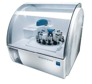
- Sample Type-Serum, plasma, whole blood, urine, CSF test can be validated
- Proteins, enzymes, lipids, hormones, etc. Levels of serum biochemical parameters, including creatinine, cholesterol, CRP, alkaline phosphates (ALP), alanine aminotransferase (ALT), total bilirubin and total protein and cytokines (interleukin 10, interleukin 4, interleukin 12, interferon gamma and tumour necrosis factor alpha) IRON, Triglyceride, Lipase urea, were measured in serum samples urine/CSF Albumin [mg/L], Haemoglobin, Bilirubin, Urea
- Below test can be performed using this equipment
- Manufacture and production of polymeric components at high volume can be done using the Injection Moulding Machine.
| ALAT (GPT) FS | CK-NAC FS | NEFA FS |
| Albumin FS | Creatinine FS | Phosphate FS |
| Alkaline Phosphatase FS | Creatinine PAP FS | Potassium FS |
| Amylase FS | Gamma GT FS | Sodium FS |
| ASAT (GOT) FS | Glucose Hexokinase FS | T4* |
| Bicarbonate FS | beta-Hydroxybutyrate FS | Total Protein FS |
| Bilirubin Auto Total FS | Iron FS Ferene | Triglycerides FS |
| Bilirubin Auto Direct FS | Lactate FS | UIBC FS |
| Calcium P FS | LDH FS | Urea FS (= BUN) |
| Chloride 21 FS | Lipase DC FS | Uric acid FS |
| Cholesterol FS | Magnesium XL FS |
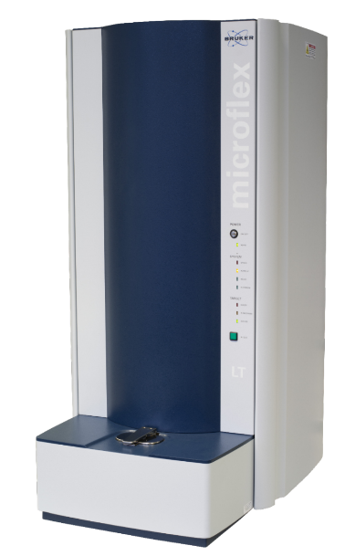
- MALDI Imaging is a powerful mass spectrometry tool for mapping the distribution of molecules from a thin sample, ranging from small metabolites to large proteins, without molecular tags or labels.
- MALDI-TOF MS is magnified by virtue of its low sample volume requirements and broad sample content (salts, buffers) amenability.
- MALDI coupled to time-of-flight mass spectrometry (MALDI-TOF MS) is used to sequence proteins, map biomolecules in tissues, identify microorganisms, and analyze several thousand biochemical assays in a day.
- This method identifies various microorganisms such as bacteria, fungi, parasites, and viruses, which supply comprehensive information. One of the MALDI-TOF MS's crucial applications is bacteriology, which helps identify bacterial species, identify toxins, and study bacterial antibiotic resistance.
- Because of its ability to perform molecular analyses while retaining morphological information, MALDI Imaging is very well suited to study molecular distribution patterns within a tumour.
- LDI-TOF MS has various benefits over the conventional method of biochemical identification, including ease of use, speed, accuracy, and low cost.
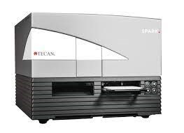
- It gives you the ability to record the whole well area of a 96 or 384 well microplate with just one image, without tiling or distortion. This means that you never miss a cell when investigating the total cell population in a microplate well. it can be used to perform following test
a. Reading Elisa’s
b. Low-volume DNA/RNA quantification
c. Nucleic acid labelling efficiency
d. Protein quantification
e. Reporter gene assays
f. Homogeneous time-resolved fluorescence (HTRF®)
g. Transcreener
h. Dual-Luciferase® Reporter (DLR™)
i. Bioluminescence resonance energy transfer (BRET) – including Nano BRET™
j. Cell counting and viability
k. Confluence assessments
l. Cell migration and wound healing
m. Fluorescence* imaging for – nuclei counting – transfection efficiency – cell viability and apoptosis – cell roughness